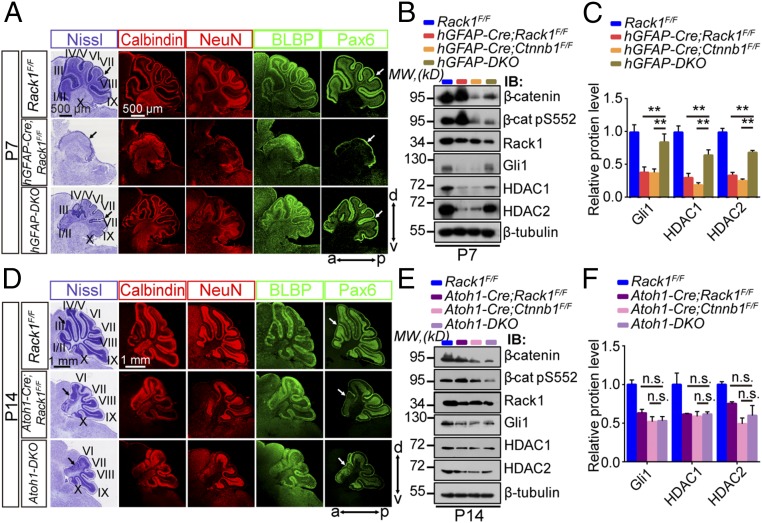
Fig. 6.
Simultaneous deletion of β-catenin in NSCs, but not GNPs, significantly rescues the Rack1 mutant cerebellar phenotype. (A and D) Nissl and immunofluorescent staining of cerebellar sections for the indicated genotypes with anticalbindin, anti-NeuN, anti-BLBP, and anti-Pax6 antibodies at P7 and P14, respectively. Arrows point to cerebellar lobules. (Scale bars: 500 μm and 1 mm.) (B) Representative Western blots examining the expression of β-catenin, Rack1, and Shh signaling downstream molecules in the control, hGFAP-Cre;Rack1F/F, hGFAP-Cre;Ctnnb1F/F, and hGFAP-DKO mutant cerebellum, respectively, at P7. IB, immunoblot; MW, molecular weight. (C) Quantitative analysis indicates significantly decreased expression of Gli1 and HDAC1/HDAC2 in both hGFAP-Cre;Rack1F/F and hGFAP-Cre;Ctnnb1F/F single-mutant mice compared with Rack1F/F controls, which could be significantly rescued in hGFAP-DKO mice (mean ± SEM; **P < 0.001, n = 4). (E) Representative Western blots examining the expression of β-catenin, Rack1, and Shh signaling downstream molecules in control, Atoh1-Cre;Rack1F/F, Atoh1-Cre;Ctnnb1F/F, and Atoh1-DKO double-mutant cerebellum, respectively, at P14. (F) Quantitative analysis indicates significantly decreased expression of Gli1 and HDAC1/HDAC2 in Atoh1-Cre;Rack1F/F, Atoh1-Cre;Ctnnb1F/F, and Atoh1-DKO muta.9nt mice compared with Rack1F/F controls. The expression of Gli1 and HDAC1/HDAC2 in Atoh1-DKO double-mutant mice is indistinguishable compared with Atoh1-Cre;Rack1F/F or Atoh1-Cre;Ctnnb1F/F single-mutant mice (mean ± SEM; P > 0.05, n = 5). n.s., not significant.

