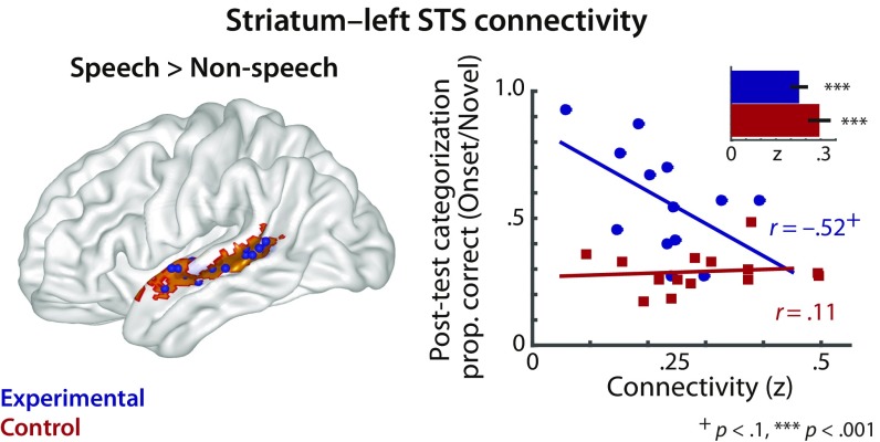
Fig. 5.
Functional connectivity between the posterior striatum and speech-selective left superior temporal sulcus tissue. (Left) l-STS tissue was identified as exhibiting greater BOLD activation for hearing speech compared with nonspeech sounds (i.e., speech: English words and syllables vs. nonspeech: semantically matched environmental sounds and sound exemplars from the videogame) in a separate localizer task before videogame training. The group-based speech > nonspeech contrast mask is shown in orange, and individually defined speech-selective ROIs are shown as blue spheres. (Right) Correlations between generalization of onset-category learning in the posttest and the extent of functional connectivity between the striatum to the localized l-STS ROIs on a single-subject basis. Behavioral categorization performance (y axis) represents generalization performance for categorizing novel onset-category exemplars. The bar graph (Inset) shows average connectivity for each group from the striatal seeds to individually defined speech-selective ROIs. The functional connectivity measure is expressed in Fisher’s z-correlation coefficient. The correlation r values are derived from post hoc correlation analyses, following upon a significant Group × Striatal Connectivity interaction effect from a multiple linear regression analysis. Error bars indicate ±1 SEM.

