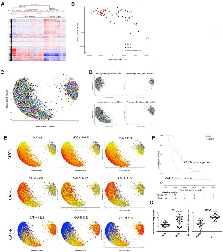
Figure 1.
The prognostic significance of the cancer-associated fibroblast (CAF) gene expression profile in ovarian cancer. A) Laser microdissection was performed on isolated epithelial tumor cells and stromal CAFs from frozen tissue samples obtained from patients with high-grade serous ovarian cancer (HGSOC; n = 70). As undifferentiated bone marrow–derived mesenchymal stem cells (MSCs) that have migrated to the tumor site have been shown to be a source of CAFs, transcriptome profiling was performed on primary bone marrow MSCs isolated from five healthy individuals. Hierarchical clustering analysis of transcriptome data generated from laser-microdissected CAFs and purified bone marrow MSCs identified two major CAF subtypes. B) Principal component analysis (PCA) on microdissected transcriptome profiles. C) With a fold change of 2 set as the cutoff, probe sets representing differentially expressed genes were visualized on the PCA loading plot. D) According to their expression levels, up- and downregulated genes in CAF-C, CAF-N, and MSCs were colored in red and blue, respectively. E) Differentially expressed probe sets represented in the PCA loading plots in three representative samples for each of the gene signature subtypes. F) The overall survival durations of patients with the CAF-C and CAF-N gene expression signatures (n = 46) were evaluated by Kaplan-Meier analysis and log-rank test. G) Transcriptome analysis of the expression levels of FAP and ACTA2 in CAFs with CAF-C and CAF-N gene expression signatures. CAF = cancer-associated fibroblast; MSC = mesenchymal stem cell.

