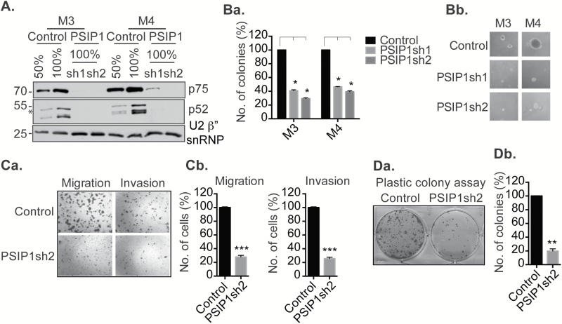
Figure 2.
PSIP1 enhances the aggressive properties of TNBC cells. (A) Western blot showing the efficiency of shRNA mediated knockdown of PSIP1 (both p75 and p52 isoforms) in M3 and M4 cells. U2β” snRNP is used as loading control. (Ba and Bb) M3 and M4 cells depleted of PSIP1 showing reduction in colony number and size in soft agar anchorage-independent colony formation assay. Colonies are counted from three independent experiments. (Ca and Cb) M4 cells depleted of PSIP1 showing reduction in migration and invasion. Cells are stained and counted from three independent experiments. (Da and Db) M4 cells depleted of PSIP1 showing reduction in long term cell proliferation, assayed by anchorage-dependent plastic colony formation assay. Cells are stained and counted from three independent experiments. Error bars in the graphs represent SEM. *P < 0.05, **P < 0.01, ***P < 0.001.

