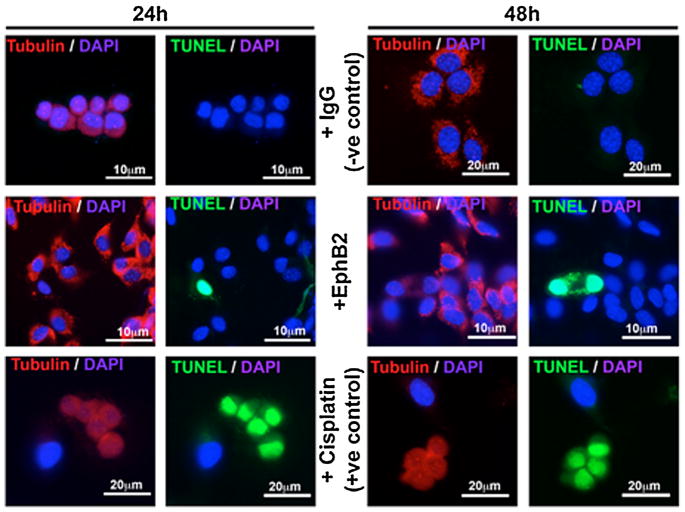
Fig. 3.
MEE cells were isolated from the mouse palatal MES and grown to 80% confluence, then treated with 5 μg/mL IgG Fc (−ve control) or clustered EphB2/Fc. At 24 or 48 h, cells were labeled with anti-tubulin (Alexa Fluor 488, red), avidin-FITC for TUNEL (green), and DAPI for nuclei (blue). IgG Fc-treated MEE showed strong tubulin expression without any TUNEL signal throughout the experiment. EphB2/Fc-treated cells showed no substantial change in Tubulin expression, and very few cells undergoing apoptosis at either 24 or 48 h. Cisplatin treated MEE cells (+ve control) underwent apoptosis within 24 h with reduced tubulin on average, and showing a characteristic rounded and clumped morphology.

