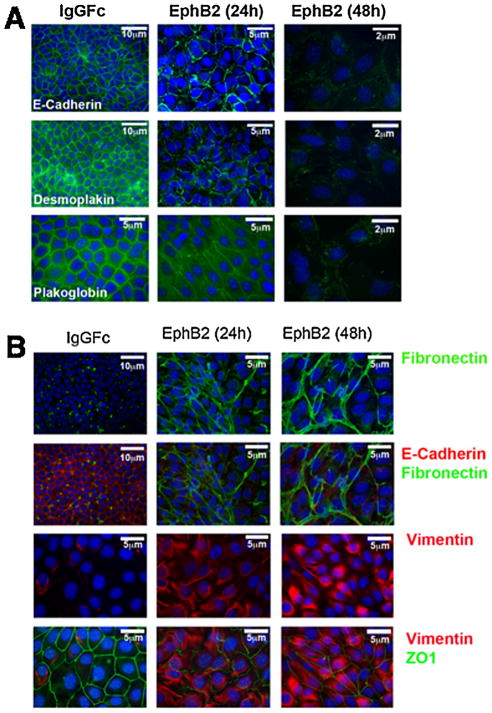
Fig. 5.
Ephrin reverse signaling causes EMT-like marker changes in mouse palatal MEE cells. Embryonic mouse MEE cells were cultured for 48 h in either IgG Fc or EphB2/Fc protein at 5 ng/mL, then fixed and processed for immunofluorescent detection of epithelial or mesenchymal markers. (A) Expression of the epithelia-specific cell junction markers E-cadherin, demosplakin, and plakoglobin (green) virtually disappeared after 48 h of EphB2/Fc treatment. (B) Expression of the mesenchymal markers fibronectin (green) and vimentin (red) increased dramatically after 48 h of EphB2/Fc exposure while expression of epithelia-associated proteins E-cadherin (red) and Z01 (green) essentially disappeared.

