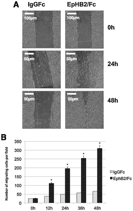
Fig. 6.
Ephrin reverse signaling induces migration of mouse palatal MEE cells. (A) Embryonic MEE cells were grown to confluence and then scratched with a needle to create a cleared area with uniform borders. The cells were treated with IgG Fc or EphB2/Fc for 48 h. (B) The number of cells that migrated across an 8 μm membrane in a transwell chamber was counted at 24 and 48 h. The change in the number of migrating cells was determined by comparison to control (IgG Fc) and plotted as numbers of migrating cells (mean ± SD.; n =3; *P <0.005 compared with controls AP-value of ≤0.05 was considered significant. The one-way ANOVA indicated that the values differ significantly across the treatment groups. All EphB2 treatment (time dependent) differed significantly (*P ≤0.005) from the control groups (IgG Fc).

