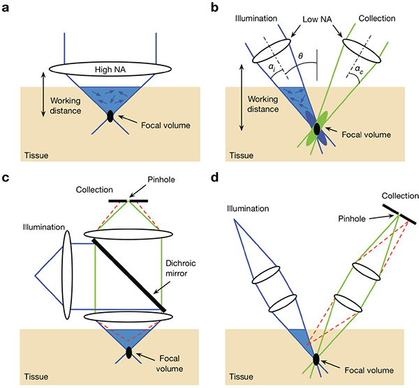
Fig. 1.

Comparison of the optical configurations for a conventional singleaxis confocal (SAC) microscope and a dual-axis confocal (DAC) microscope, (a) In order to achieve a tight focal volume (black oval), a SAC microscope requires a high-N A objective lens. This results in a short working distance that makes miniaturization and beam scanning more difficult, (b) A DAC microscope uses low-NA off-axis illumination and collection beams, in which the focal volume is defined by the overlapping foci of the two beams. The use of low-NA beams allows for a longer working distance, which provides advantages for miniaturization and beam scanning, (c) In SAC microscopy, out-of-focus light (an example beam path is shown with the dashed red lines) is not completely rejected by the pinhole, (d) In DAC microscopy, out-of-focus light is directed away from the pinhole and is more optimally rejected, thereby improving the signal-to-background ratio.
