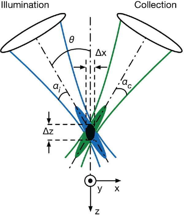
Fig. 2.

The DAC microscope architecture. Two low-NA beams (illumination and collection) with focusing angles of αi and αc, respectively, intersect at a half-angle of θ. The focal volume (black oval) of the system is defined by the product of the intersecting point spread functions (PSFs) of the illumination (blue) and collection (green) beams. The dimensions of the focal volume (ᐃx, ᐃy, and ᐃz) correspond to the spatial (lateral and axial) resolutions of the system.
