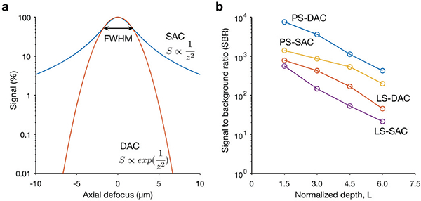
Fig. 3.

Quantitative comparison of the axial-sectioning response (a) and contrast (b) of typical SAC and DAC systems, (a) The theoretical axial response of a SAC and DAC microscope is shown, in which a point object is translated through the focus of the microscope in the z direction. In this case, the SAC and DAC microscopes have equivalent axial resolutions (FWHM). The signal rolls off more quickly in a DAC system (red) than in a SAC system (blue), showing that the axial sectioning performance (rejection of out-of-focus light) is superior for the DAC configuration. (b) Monte-Carlo scattering simulations to compare the contrast (signal-to-background ratio, SBR) of various microscope configurations when imaging an in-focus reflective object within highly scattering biological tissues, as a function of depth. The results show that both the point-scanned (PS) and the line-scanned (LS) versions of DAC microscopy provide superior image contrast in scattering media when compared with their SAC microscopy counterparts. The normalized depth refers to the number of mean free paths that ballistic photons would travel in a round-trip perpendicular path from the tissue surface to the focal volume, i.e. L=2μsd, where μs is the scattering coefficient, and d is the imaging depth in the direction normal to the tissue surface [46]
