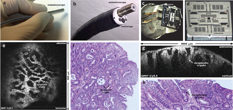
Fig. 5.

Photographs of a miniature DAC endo-microscope (a) that is fitted within a clinical GI endoscope (b). The head of the imaging probe has a diameter of 5.5 μm. (c-d) Photograph and scanning electron micrograph (SEM) of a custom-developed tri-axial MEMS scanner that enables the user to switch between two orthogonal imaging planes (either the en face plane or the vertical plane) in real time. (e) An example of en face optical sectioning of mouse colon after intravenous injection of a Cy5.5-labeled peptide, showing the dysplastic crypts (arrows) and goblet cells (arrowheads). (f) A corresponding H&E-stained histology section is shown of the mouse colon. (g) An example of vertical optical sectioning of the same mouse colon, showing EGFR expression from a region of adenoma up to 430 μm below the surface. (h) A corresponding H&E-stained histology section is shown of the mouse colon. Ref: [74]
