
Fig. 6.
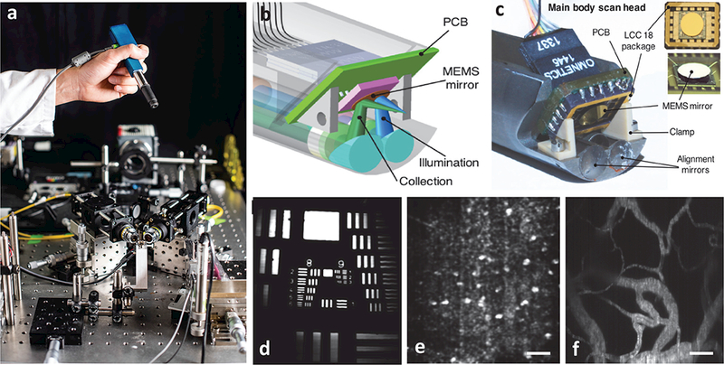
A handheld video-rate LS-DAC microscope for intraoperative guidance. (a) A miniature LS-DAC is held above a large tabletop LS-DAC prototype [photograph courtesy of Dennis Wise at the University of Washington]. (b-c) A design rendering and photograph, respectively, of the scan head of the handheld LS-DAC device. A single MEMS mirror is used to scan both beams in one dimension to rapidly generate 2D images. (d) An image of a 1951 USAF resolution target, showing the ability to resolve objects as small as ~1 μm. (e) A label-free in vivo image of hyper-reflective nuclei in human oral buccal mucosa. (f) A fluorescence in vivo image (maximum intensity projection from a depth range of 50 to 100 μm) of the vasculature of a mouse ear after retro-orbital injection of FITC-dextran. All scale bars represent 50 μm.