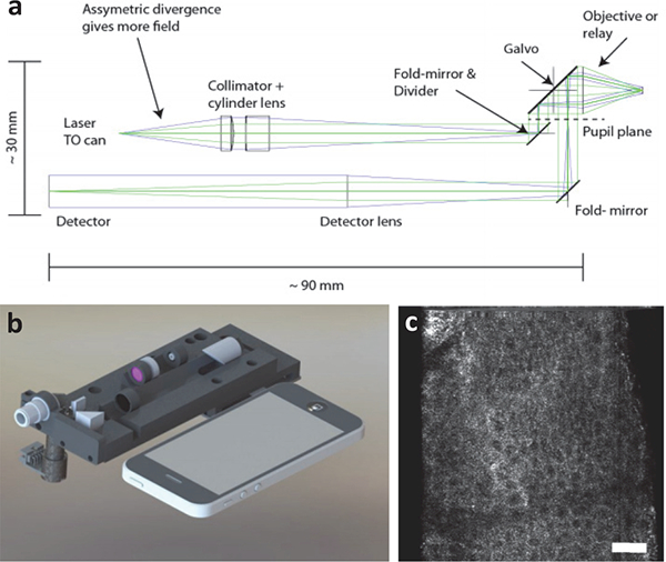
Fig. 7.

(a) Optical circuit of a compact line-scanned divided pupil confocal system. The pupil of the objective lens is physically divided into two halves, one to generate the illumination beam and the other for the collection path. A single galvanometric mirror is used to scan both beams to create an image. (b) A design rendering of the system is shown alongside a smartphone. (c) A label-free in vivo image is shown of human epidermis. Ref: [76]
