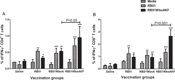
Fig 9.
Flow cytometric analysis showing the percentage of interferon-γ secreting (A) CD4+ and (B) CD8+ T cells in the spleens of immunized mice. Mice were immunized intra-peritoneally with 108 CFU of RB51, RB51WboA, RB51WboAKF, or inoculated with saline. Their splenocytes were harvested and stimulated in vitro with media (unstimulated), heat-killed RB51 or RB51WboAKF. Values are shown as mean ± standard deviation (n = 4). Asterisks indicate statistically significant differences from the corresponding unstimulated control. *, P < 0.05; **, P < 0.01; ***, P < 0.001.

