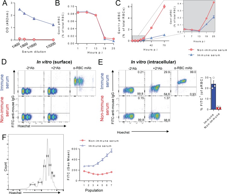
Fig 8. Antibodies induced by P. chabaudi chabaudi AS infection, also protect by blocking parasite transit between RBC, not accelerating pRBC clearance.
(A) Assessment of PcAS-specific total IgG in diluted sera from infected or age-matched naïve mice (n = 10/group), taken at day 40 p.i. with PcAS. (B-C). Mice (n = 5/group) were injected with PcAS-immune serum or non-immune control serum 24h prior to challenge with CTFR+ PcAS-infected pRBCs, with peripheral blood monitored at times indicated for: (B) loss of Gen0 (CTFR+) pRBC over the first 25h, and (C) emergence of Gen1+ pRBC over 73h, with the zoomed-in box showing the first 25h in more detail. (D-E) Assessment of in vitro deposition of mouse IgG from PcAS-immune or non-immune control serum performed in triplicate, either (D) on the surface of RBC from PcAS-infected mice, or (E) inside RBC from PcAS-infected mice after fixation/permeabilisation. Graph shows percentage of pRBC binding to mouse IgG after fixation/permeabilisation. (F) Representative histogram shows gating for identifying PcAS-infected pRBC containing different amounts of parasite DNA using Hoechst. Graph shows variation in geometric mean fluorescence intensity of in vitro IgG deposition inside fixed and permeabilised pRBC according to DNA content, when incubated with immune or non-immune control serum (assessed in triplicate). Data in (D-F) are representative of two independent experiments each with similar results.

