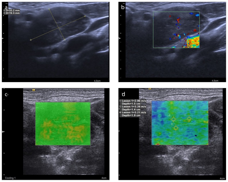
Figure 4. a–d.
A 49-year-old man with Hodgkin lymphoma. B-mode US image (a) reveals a 33.2×19.3 mm lymph node with heterogeneous internal structure, loss of echogenic hilus, and 0.58 axial ratio at level 3. Abnormal mixed type vascularity pattern is seen on color Doppler (b). The VTIQ quality map (c) is green, indicating decent VTIQ estimates. VTIQ velocity mode image (d) shows the SWVs (m/s) of three different points. Histopathologic diagnosis was made after excisional lymph node biopsy because the lymph node could not be diagnosed by fine-needle aspiration biopsy.

