Figure 3.
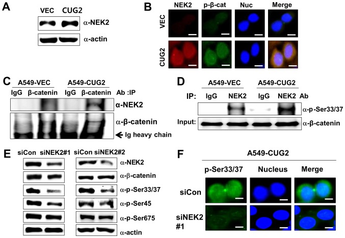
CUG2-activated NEK2 is responsible for the phosphorylation of β-catenin at Ser33/Ser37. (A) Lysates of A549-VEC and A549-CUG2 cells were separated by performing SDS-PAGE on 10% gels, and NEK2 was detected by performing western blot analysis with anti-NEK2 antibody. (B) Levels of NEK2 and β-catenin phosphorylated at Ser33/Ser37 (p-β-catenin; p-Ser33/Ser37) in A549-VEC and A549-CUG2 cells were determined by performing immunofluorescence microscopy with Alexa Fluor 594-conjugated goat anti-mouse IgG (red) and Alexa Fluor 488-conjugated goat anti-rabbit IgG (green), respectively. For performing nuclear staining, DAPI was added before mounting in glycerol. Scale bar indicates 10 µm. (C) β-catenin was pulled down from the lysates of A549-VEC and A549-CUG2 cells by using an anti-β-catenin antibody. NEK2 present in the immunoprecipitates was detected using an anti-NEK2 antibody, and β-catenin present in the immunoprecipitates was detected as a loading control by using an anti-β-catenin antibody. (D) NEK2 was pulled down from A549-VEC and A549-CUG2 cells by using an anti-NEK2 antibody or isotype IgG as a control. The reaction mixture for the NEK2 kinase assay, including immunoprecipitates as NEK2 kinase, recombinant GST-β-catenin as a substrate, ATP, and a reaction buffer, was incubated at 30°C for 1 h. NEK2 kinase activity was analyzed by determining the phosphorylation of GST-β-catenin at Ser33/Ser37 by performing immunoblotting, and β-catenin levels were examined as a loading control. (E) Lysates of A549-CUG2 cells transfected with the control or NEK2 siRNAs (#1 and #2) were separated by performing SDS-PAGE on 10% gels. Phosphorylation states of β-catenin following treatment were detected using antibodies against Ser33/Ser37/Thr41, Ser45 and Ser675 of β-catenin. (F) Following transfection with control or NEK2 siRNA#1, the levels of p-β-catenin (p-Ser33/Ser37) in A549-CUG2 cells were detected by performing immunofluorescence microscopy with Alexa Fluor 488-conjugated goat anti-rabbit IgG (green). For performing nuclear staining, DAPI was added before mounting in glycerol. Scale bar indicates 10 µm. CUG2, cancer-upregulated gene 2; NEK2, never in mitosis gene A-related kinase 2.

