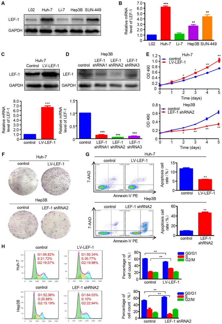
Figure 4.
LEF-1 enhances cell proliferation and inhibits apoptosis and cell cycle arrest in HCC cells. (A) Western blotting was used to detect the LEF-1 protein expression in the HCC cell lines Huh-7, Li-7, Hep3B and SNU-449, and the normal hepatic cell line L02. (B) RT-qPCR was used to detect the LEF-1 mRNA expression in the HCC cell lines Huh-7, Li-7, Hep3B and SUN-449, and the normal hepatic cell line L02 (n=3; **P<0.01, ***P<0.001 vs. L02). (C) The protein and mRNA expression of LEF-1 was evaluated in the Huh-7 cells transfected with LV-LEF-1 using western blotting and RT-qPCR analysis, respectively (n=3; ***P<0.001 vs. control). (D) The protein and mRNA expression of LEF-1 was detected in the Hep3B cells transfected with three LEF-1 shRNAs using western blotting and RT-qPCR analysis, respectively (n=3; ***P<0.001 vs. control). (E) MTT assay was performed to detect the proliferation of Huh-7 cells transfected with LV-LEF-1 and Hep3B cells transfected with LEF-1 shRNA (n=3; *P<0.05, **P<0.01 vs. control). (F) The colony-forming ability was assessed using the colony formation assay. Representative images are shown. (G) Flow cytometric analysis was used to observe the apoptosis rate of Huh-7 and Hep3B cells following transfection with LV-LEF-1 or LEF-1 shRNA, respectively (n=3; **P<0.01 vs. control). (H) The DNA content was analyzed in Huh-7 and Hep3B cells transfected with LV-LEF-1 or LEF-1 shRNA using flow cytometry, and the percentage of cells in the G0/G1, S and G2/M phases of the cell cycle was calculated (n=3; **P<0.01). HCC, hepatocellular carcinoma; RT-qPCR, reverse transcription-quantitative polymerase chain reaction; LEF-1, lymphoid enhancer-binding factor 1; LV, lentivirus; shRNA, short hairpin RNA: 7-AAD, 7-aminoactinomycin D; PE, phycoerythrin.

