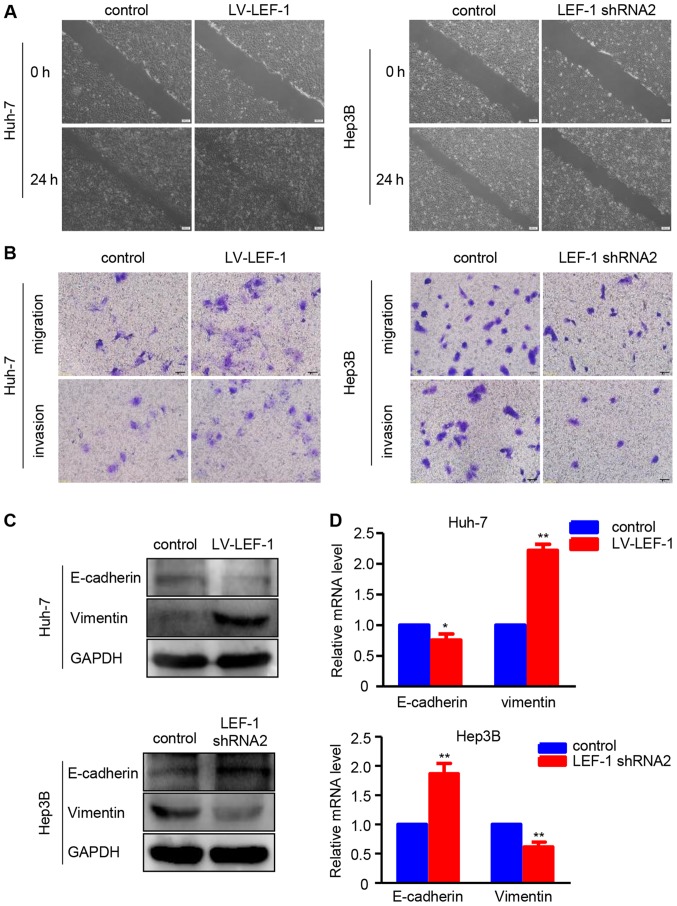
Figure 5.
LEF-1 promotes migration, invasion and epithelial-to-mesenchymal transition in hepatocellular carcinoma cells. (A) The wound healing assay was performed to observe the cell migration ability at 24 h after transfection with LV-LEF-1 or LEF-1 shRNA in Huh-7 and Hep3B cells. Representative images are shown. Scale bars, 200 µm. (B) The Transwell migration assay and Transwell Matrigel invasion assay were performed to examine the migration and invasion ability of Huh-7 and Hep3B cells following transfection with LV-LEF-1 or LEF-1 shRNA. Representative images are shown. Scale bars, 50 µm. (C) The protein expression of E-cadherin and vimentin was detected in Huh-7 and Hep3B cells following transfection with LV-LEF-1or LEF-1 shRNA using western blot analysis. (D) The mRNA expression of E-cadherin and vimentin was detected in Huh-7 and Hep3B cells following transfection with LV-LEF-1 or LEF-1 shRNA using reverse transcription-quantitative polymerase chain reaction. (n=3; *P<0.05, **P<0.01 vs. control). LV, lentivirus; LEF-1, lymphoid enhancer-binding factor 1; shRNA, short hairpin RNA.

