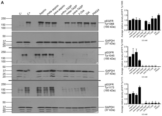
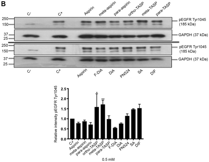
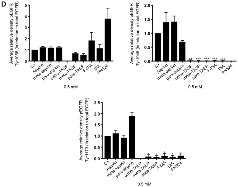
Figure 7.
Effect of aspirin and analogues on pEGFR tyrosine kinase phosphorylation sites and EGFR. SW480 colorectal cancer cells were incubated with human rEGF (200 ng/ml) for 5 min following being treated with 0.5 mM compound (HEPES buffered) for (A) 24 h and probed with anti-human EGFR pY1068, pY1045 and pY1173 antibodies accompanied by corresponding histograms, (B) 2 h and probed with anti-human EGFR pY1045 and (C) 24 h and probed with an anti-human EGFR (D38B1) XP rabbit antibody. (D) Quantification of the effect of these compounds on pEGFR tyrosine kinase phosphorylation sites 1,068, 1,045 and 1,173 in relation to total EGFR expression. Cell lysates were analysed by SDS-PAGE and immunoblotting. pEGFR and EGFR bands migrated at ~185 and ~180 kDa, respectively, molecular weight was estimated by interpolation using Precision Plus colour standards. Band intensities were quantified relative to GAPDH. C- represents unstimulated and untreated cells; C+ represents untreated cells stimulated with rEGF for 5 min. Data presented are the mean of three individual experiments ± standard error of the mean. *P<0.05, **P<0.01 and ***P<0.001 vs. C+ (one-way analysis of variance followed by Dunnett’s test). EGF, epidermal growth factor; EGFR, EGF receptor; pEGFR, phosphorylated EGFR; rEGF, recombinant EGF; Y, tyrosine; SA, salicylic acid; DIF, diflunisal.


