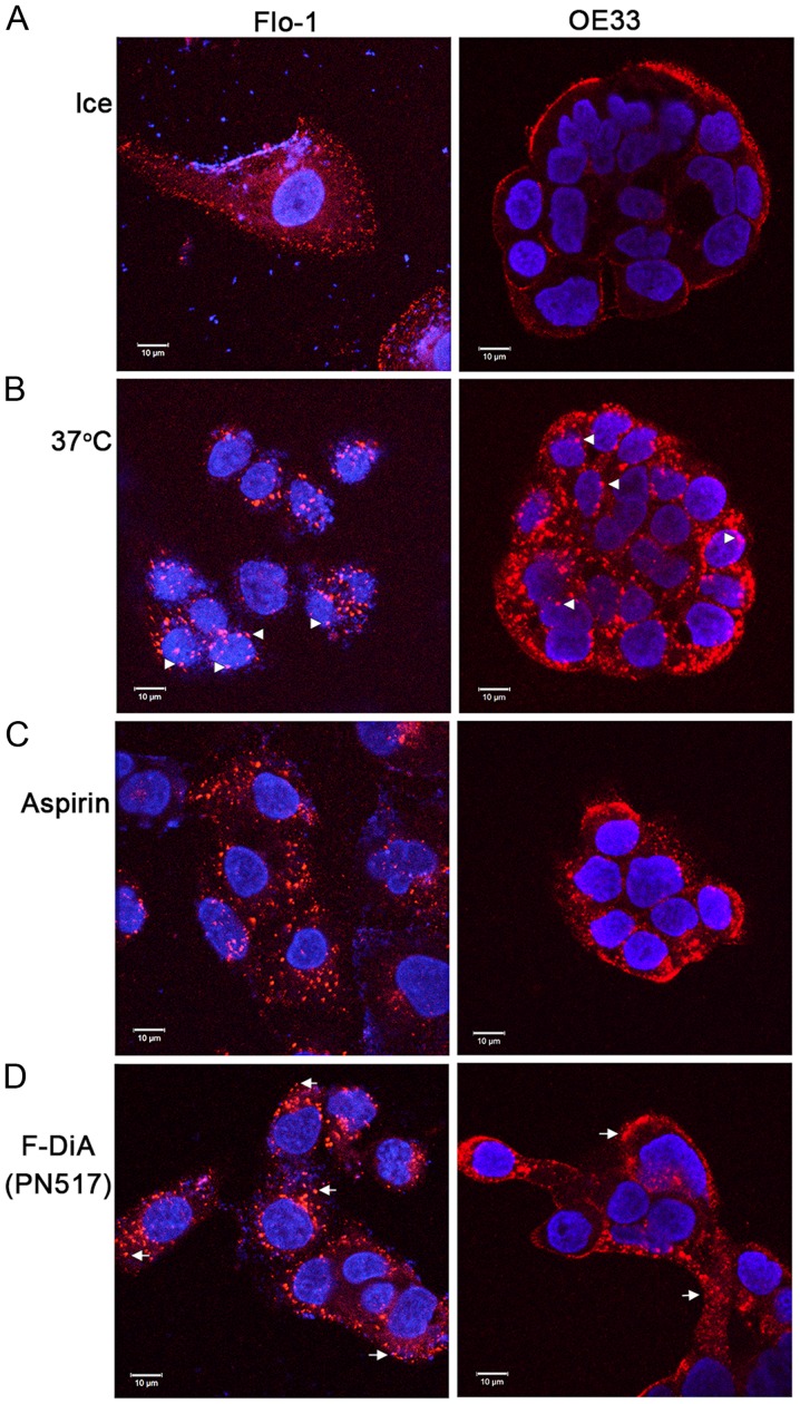
Figure 8.
Effect of aspirin analogues under HEPES-buffered conditions on EGF (20 ng/ml) internalisation in Flo-1 and OE33 oesophageal cell lines. Confocal analysis of oesophageal cancer cells incubated with (0.5 mM) or without compound and fluorescence-conjugated EGF. (A) Negative control, without EGF internalisation in the absence of compound. Cells warmed to 37°C in (B) the absence of compound and the presence of (C) aspirin and (D) F-DiA. Images were acquired at 405 nm for DAPI nucleic stain (blue) and at 561 nm for Alexa Fluor 555-EGF (red). Representative images were recorded at ×40 magnification using an oil/1.30 NA oil immersion objective lens. The close proximity of EGF towards the nucleus upon internalisation with warming is observed in Flo-1 cells (arrowheads). In cells incubated with compound the EGF is less centrally located (arrows), particularly with F-DiA in Flo-1 cells. EGF, epidermal growth factor; F-DiA, fumaryldiaspirin.

