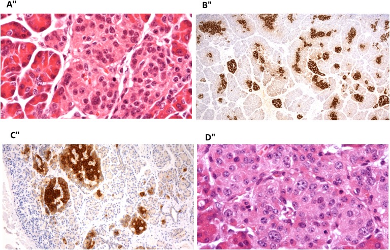
Figure 1.
Histopathology of pancreas after sirolimus use. (A) Patient with diffuse CHI resulting from a homozygous ABCC8 mutation, 4 mo after sirolimus treatment. High-power view of an islet showing scattered enlarged islet-cell nuclei. This change was present in islets throughout the tissue, in keeping with diffuse disease. (B) Patient with focal CHI resulting from paternal compound heterozygous ABCC8 mutation, 12 mo after sirolimus. There is an increase in islet-cell tissue with strong proinsulin staining. There was no diffuse islet-cell nuclear enlargement. The appearances suggest an atypical focal form of the disease. (C) Patient with focal CHI resulting from a paternal compound heterozygous ABCC8 mutation, 21 mo after sirolimus use. The image shows enlarged and clustered islets that stain strongly for proinsulin, in keeping with focal disease. (D) Patient with diffuse CHI resulting from homozygous ABCC8 mutation, 24 mo after sirolimus. There were scattered enlarged islet-cell nuclei throughout the tissue, indicating diffuse disease. Images taken with Nikon Eclipse E600 microscope; (A, D) Stained with hematoxylin and eosin; (B, C) stained with antibody to proinsulin. Original magnification: (A) ×400, (B) ×20, (C) ×100, (D) ×400.

