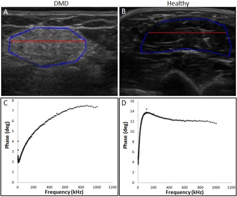
Figure 1:
Top Panel: Echo-intensity is higher in ultrasound image (GLA) of biceps brachii muscles from a 12-year-old boy with DMD (A) compared to a same-aged healthy control (B). The area within the blue line demarcates the region of interest and area above the red line is the most superficial one third. Bottom Panel: Example of multifrequency impedance spectra of biceps brachii from the same patient (C) and the healthy control (D).

