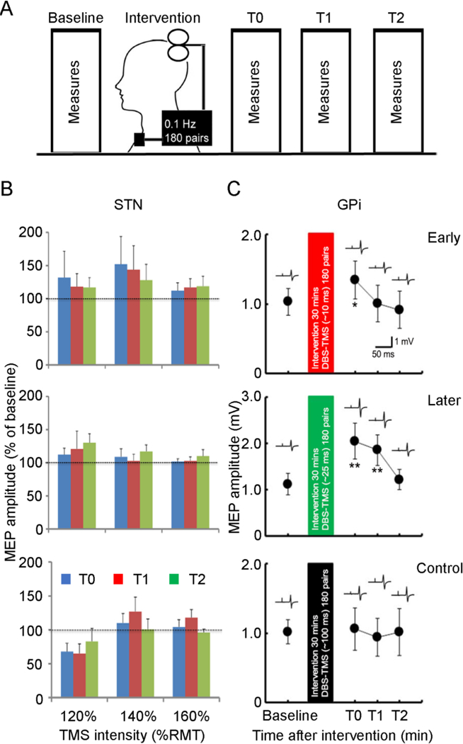
Figure 5. Induction of motor cortical plasticity by pairing deep brain stimulation with transcranial magnetic stimulation.

(A) Experimental setup. Motor cortical plasticity was tested by a paired associative stimulation paradigm. Interventional protocols with paired associative stimulation were applied with 180 pairs of DBS and TMS at 0.1 Hz for 30 min. Three different interstimulus intervals (early, later and a control interval) were tested. Measurements were performed before (baseline) and at three different time points after the interventional protocol (T0 immediately, T1 about 30 minutes, T2 about 60 minutes after the intervention). (B) and (C) Group analysis (mean ± standard deviation) for the effect on motor cortical plasticity in Parkinson’s disease patients with STN DBS (B) and in cervical dystonia patients with GPi DBS (C). (B) Interstimulus intervals of 3 ms (early), 23 ms (later) and 167 ms (control) were tested in patients with STN DBS. MEP recruitment curve were measured with TMS intensity set at 120%, 140%, and 160% of RMT. The abscissa indicates TMS intensities. The ordinate indicates MEP amplitudes normalized as a percentage of that before intervention (baseline). Blue columns represent time T0, red columns represent time T1, green columns represent time T2. Analysis of variance revealed a trend toward significance for the interaction between interventional protocol and TMS intensity (P=0.07). (C) Interstimulus intervals of ~10 ms (early), ~25 ms (later) and 100 ms (control) were tested in patients with GPi DBS. The abscissa indicates the time after interventional protocol. The ordinate indicates MEP amplitude. Example of recordings shows MEP measured at the corresponding time points. Paired associative stimulation with interstimulus interval of ~25 ms produced MEP facilitation for longer than 30 minutes (note the different ordinate for this panel) and that with interval of ~10 ms produced MEP facilitation at the time point immediately after the intervention. MEP after the paired associative stimulation with interstimulus interval of 100 ms did not change. *P<0.05, ** P<0.01, post hoc t-test, comparing MEP after paired associative stimulation to that at baseline. DBS = deep brain stimulation, GPi = internal globus pallidus, MEP = motor evoked potential, RMT = resting motor threshold, STN = subthalamic nucleus, TMS = transcranial magnetic stimulation. Modified from Udupa et al. (2016) and Ni et al. (2018).
