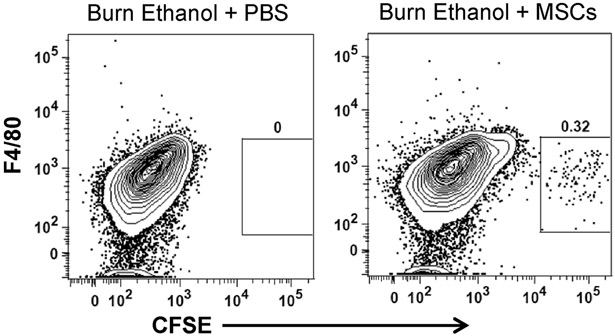
Fig 1.
Flow cytometry of MSCs in dissociated lung tissue. Representative flow cytometry plots of each treatment group. CFSE+ MSCs were identified in dissociated lung tissue from intoxicated and injured mice at 24 h using flow cytometry. Total dissociated lung cells were negatively selected for Siglec-F and CD11b (data not shown), followed by F4/80− CFSE+ cells (box) in burn ethanol + PBS and burn ethanol + MSCs treatment groups.

