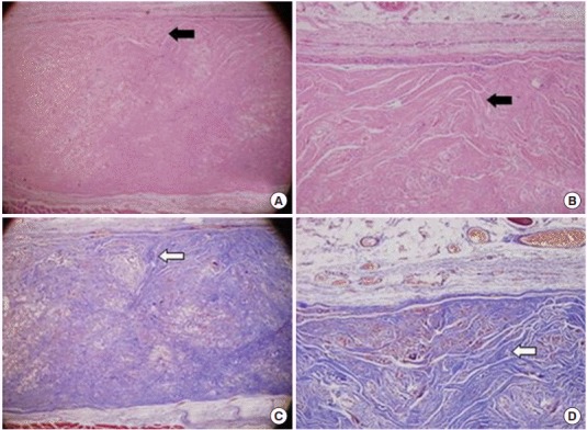
Fig. 3.

Representative photographs of grafted human acellular dermal graft (Megaderm) at 12 weeks. (A, B) A grafted acellular cadaveric dermal graft showed diffused fibrocyte like cells and borderline form between epithelium (black arrows; H&E). (C, D) Extremely high density of collagenous fibers look like a normal dermis tissue with Masson’s trichrome staining (white arrows). (A, C) ×40, (B, D) ×100.
