Figure 1.

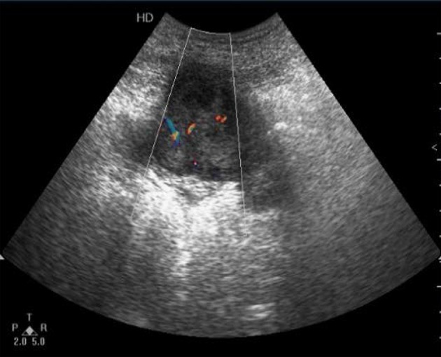
Detection of the right lower abdomen. Irregular hypoechoic mass which showed lobulated enhanced echoes in the surrounding intestine and omentum, and enriched internal blood flow signals within the mass.

Detection of the right lower abdomen. Irregular hypoechoic mass which showed lobulated enhanced echoes in the surrounding intestine and omentum, and enriched internal blood flow signals within the mass.