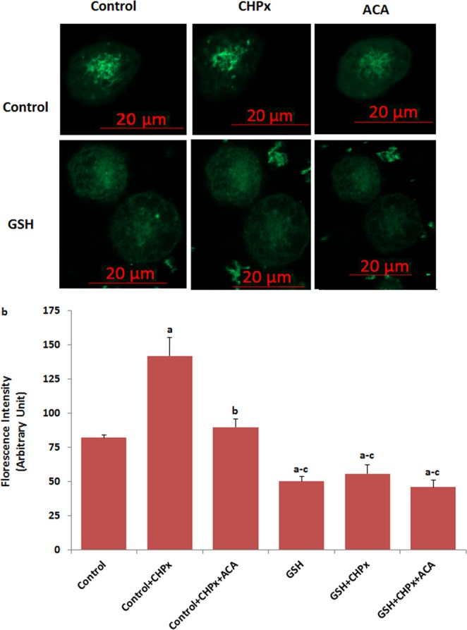
Figure 1.

Activation of TRPM8 in the Du 145M8 cells by oxidative stress. (mean ± SD). The cells were stained with Fluo-3 calcium dye and mean ± SD of fluorescence in 15 mm2 of cell as arbitrary unit are presented; n = 10–20 independent experiments. In GSH experiments, the cells were pretreated with GSH (10 mM for 2 hours). The cells were extracellularly stimulated by cumene hyroperoxide (CHPx and 1 mM for 5 min) but they were extracellularly inhibited by ACA (25 μM for 10 min). The samples were analyzed by the laser confocal microscopy fitted with a 40× oil objective. The scale bar was 20 µm. Representative images and fluorescence intensities of the CHPx, ACA and GSH effect on the TRPM8 activation in the laser confocal microscope analyses are shown in (a,b) respectively. (ap ≤ 0.001 versus control. bp ≤ 0.001 versus control + CHPx group. cp ≤ 0.001 versus control + CHPx + ACA group).
