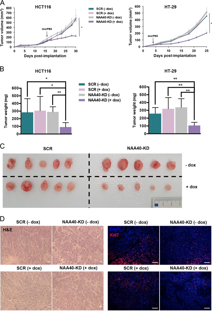
Fig. 4. NAA40 depletion inhibits CRC cell growth in vivo.
a The mean volume of tumors from the dox-treated (+dox) and PBS-treated (−dox) HCT116 (left) or HT-29 (right) xenografts harboring the conditionally induced SCR or NAA40-KD2 shRNAs. The tumor volume is shown as mean ± s.d. Unpaired two-tailed Student’s t-test was used (*p < 0.05). b At the end of the experiment in (a) mice were sacrificed and tumors were excised and weighted. Error bars represent mean ± s.d. Unpaired two-tailed Student’s t-test was used (*p < 0.05, **p < 0.01). c Representative tumors excised from SCR and NAA40-KD mouse xenografts after administration of dox (+dox) or PBS (−dox). d Representative hematoxylin & eosin (H&E) images from tumors derived from SCR and NAA40-KD mouse xenografts after administration of dox (+dox) or PBS (−dox). Scale bar, 200 μm. Representative immunofluorescence Ki67 staining (red) images from tumors derived from SCR and NAA40-KD mouse xenografts after administration of dox (+dox) or PBS (−dox). Cell nuclei were visualized using DAPI staining (blue). Scale bar, 100 μm

