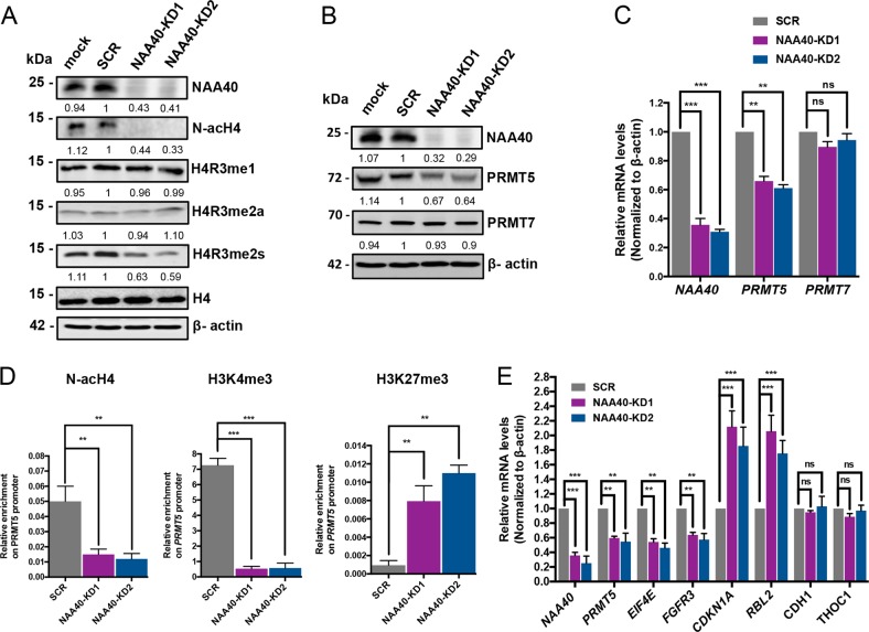
Fig. 5. NAA40 regulates PRMT5 expression.
a Western blot analysis of protein extracts from the indicated transiently transfected HCT116 cells using antibodies against NAA40, H4R3me1, H4R3me2a, H4R3me2s, total histone H4, and β-actin as loading control. The densitometry numbers below each blot define the normalized levels of H4R3me1, H4R3me2a, and H4R3me2s against H4 and of NAA40 against β-actin relative to SCR cells. b Western blot analysis of protein extracts from the indicated siRNA-treated cells using antibodies toward NAA40, PRMT5, PRMT7, and β-actin as loading control. The values below each blot were calculated by densitometry analysis of NAA40, PRMT5, and PRMT7 bands relative to SCR control after normalization with β-actin. c qRT-PCR analysis of mRNA levels of NAA40, PRMT5, and PRMT7 normalized to β-actin in the indicated HCT116 cells. Error bars represent the mean ± s.d of three biological replicates. Unpaired two-tailed Student’s t-test was used (ns = no significance, **p < 0.01, ***p < 0.001). d ChIP assay monitoring the enrichment of N-acH4, H3K4me3, and H3K27me3 on the PRMT5 gene promoter. The enrichment from each antibody was normalized to total histone H3. Results are shown as mean ± s.d. from two independent experiments. Unpaired two-tailed Student’s t-test was used (**p < 0.01, ***p < 0.001). e qRT-PCR analysis of mRNA levels in HCT116 cells for the indicated genes. The results were normalized to β-actin mRNA and represent the average from three independent experiments. Unpaired two-tailed Student’s t-test was used (ns = no significance, **p < 0.01, ***p < 0.001)

