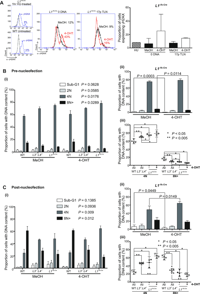
Figure 3.
LIG1-depleted cells maintain an intact DNA damage response and suppressed mitotic slippage. (A) Flow cytometry detection of the phosphorylated (Ser139) histone variant H2AX (γH2AX; red overlays) as a surrogate marker for DSBs in G2-arrested and 4-OHT-treated L1−/fx:Cre cells transfected with 17p TLN or 0 DNA control over 24 h. The top left insert shows a histogram of γH2AX-expressing (8% population; blue overlay) unsynchronized and untreated L1−/fx:Cre cells exposed to 2 mM hydroxyurea (HU) for 1 h at 37°C. The bottom left insert reveals the basal 1.6% γH2AX-expressing asynchronous and untransfected WT HCT116 cells. The right-hand bar chart shows mean proportions (with SD) of γH2AX-expressing G2-arrested L1−/fx:Cre cells 24 h post-nucleofection (two replicas). The proportions of cells with defined DNA content in three independent experiments as assessed by DAPI DNA staining of fixed G2-arrested WT, L3−/−:LIG4−/− and L1−/fx:Cre cells (B) pre-nucleofection or (C) 24 h post-nucleofection (pooled 17p TLN and 0 DNA transfections) with detection by image cytometry. (i) Data for each fraction of pre- and post-nucleofection samples is plotted as the mean with standard deviation and assessed using one-way Kruskal–Wallis ANOVA of individual population fractions (significance set as alpha = 0.05) recorded against the colour key code. Differences relating specifically to the 4-OHT-mediated deletion of LIG1 from L1−/fx:Cre cells are shown in B(ii) and C(ii). Significance was assessed by one-tailed paired T-tests of common fractions. The significant (Mann–Whitney U-tests, 0.05; *, 0.005; **) variations in proportions of WT, L3−/−:L4−/− and L1−/fx:Cre cells treated with 4-OHT are displayed in B(iii) and C(iii).

