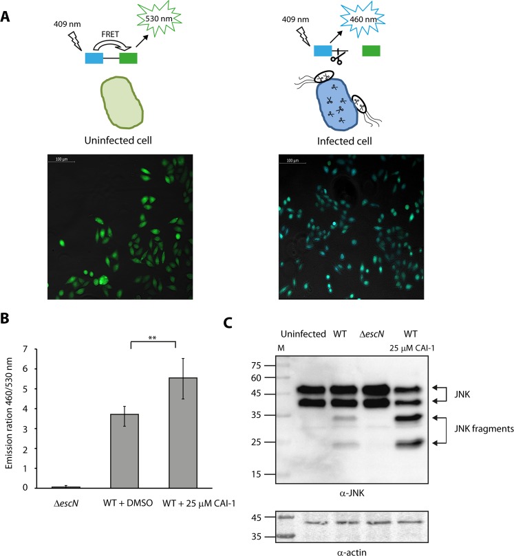
Figure 6.
CAI-1 increases EPEC translocation activity into HeLa cells. (A) Scheme of the effector translocation assay. HeLa cells loaded with CCF2‐AM emit green fluorescence (530 nm) when excited at wavelength of 409 nm. However, when the fusion protein of EspZ with β‐lactamase (EspZ-TEM) translocates into HeLa cells via the T3SS of WT EPEC, it mediates cleavage of CCF2‐AM, which induces a shift from green to blue fluorescence (460 nm). Representative images of HeLa cells either uninfected or infected with WT EPEC and loaded with CCF2-AM were imaged by fluorescence microscopy and presented as merged blue/green emission images. (B) WT EPEC carrying pEspZ-TEM was grown in the presence of 0.25% (v/v) DMSO or 25 μM CAI-1. Effector translocation levels are defined as the ratio between blue (460 nm) and green (530 nm) fluorescence. Data are the means of at least three experiments; bars represent standard error; **P < 0.005. (C) HeLa cells were infected with WT EPEC in the presence or absence of 25 μM CAI-1. After 1 h, cells were washed, and host cell proteins were extracted and subjected to western blot analysis using anti-JNK and anti-actin (loading control) antibodies. JNK and its degradation fragments are indicated at the right of the gel. WT EPEC showed degradation of JNK compared to the uninfected sample and the samples infected with ΔescN. HeLa cells infected with WT EPEC in the presence of 25 μM CAI-1 showed an enhanced JNK degradation profile compared to the WT EPEC without CAI-1, thus suggesting that CAI-1 promotes the ability of EPEC to translocate effectors into host cells.

