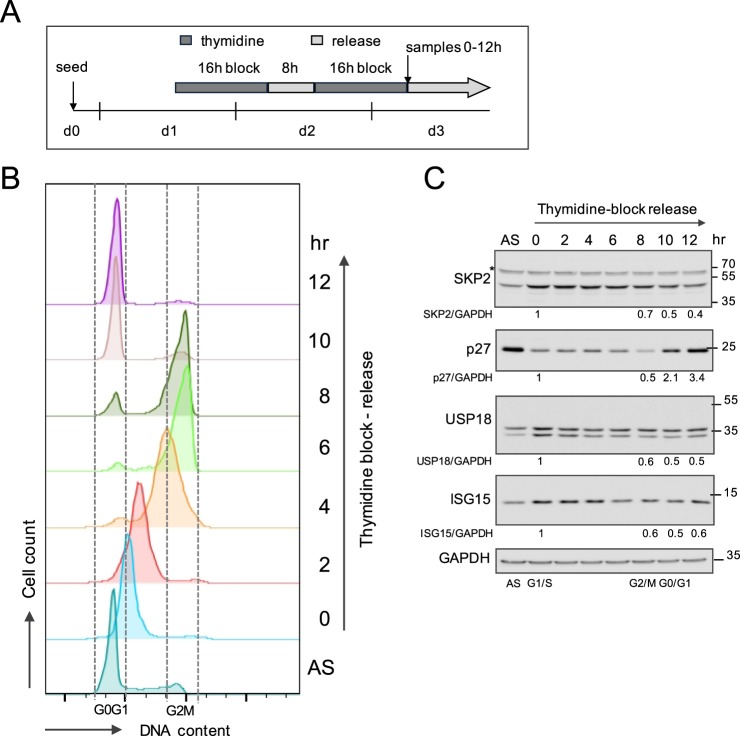
Figure 5.
Synchronized HeLa cells are analyzed for SKP2 and ISG proteins. (A) Schematics of HeLa S3 cells synchronization in G1/S arrest by double thymidine block and release into fresh medium. After the second release, samples were collected every 2 h during 12 h. (B) Cells were stained with propidium iodide for DNA content and the cell cycle profile was determined by flow cytometry. Debris and sub-G1 phase material were excluded. G0/G1 (2 N) and G2/M (4 N) phases are delineated. (C) An aliquot of cells from the samples analyzed in (B) was lysed for protein analysis. Lysates were immunoblotted, as indicated. Results are reported as the ratio of each protein to GAPDH. The ratio obtained for G1/S phase was set to 1.

