Figure 3.
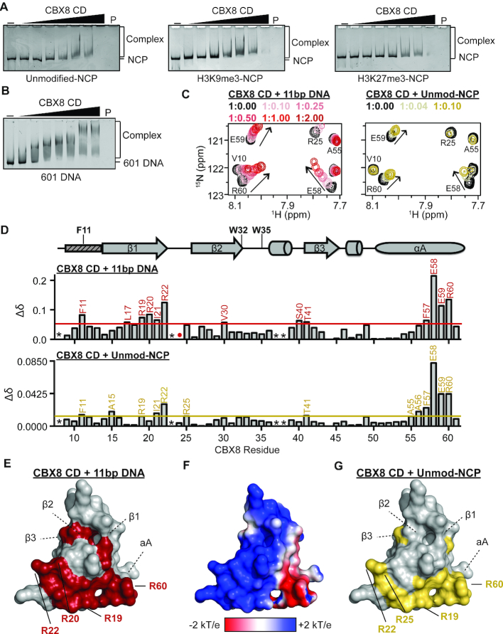
CD association with nucleosomes is driven by interactions with DNA through an arginine-rich basic patch. (A) EMSAs performed with CD and unmodified (left), H3K9me3 (middle) and H3K27me3 (right) NCPs. Shown are representative gels from a triplicate of experiments. (B) EMSAs performed with CD and the 147 bp 601 DNA. Shown is a representative gel from a triplicate of experiments. (C) 1H–15N-HSQC overlays for 15N-CD upon addition of increasing concentrations of an 11bp DNA (left) and unmodified NCP (right). Molar ratio of CD:DNA and CD:umodified NCP are color coded as shown in legend. A selected regions of the CD spectrum is shown for clarity. (D) Normalized CSP (Δδ) between the apo and DNA-bound (1:2.00 ratio) spectra are plotted against CBX8 residue number (top). Normalized CSP (Δδ) between the apo and unmodified NCP-bound (1:0.10 ratio) spectra are plotted against CBX8 residue number (bottom). CSPs were considered significant if greater than the mean plus one standard deviation and are labeled in red or gold for 11 bp DNA and unmodified NCP, respectively. The secondary structure of CD from the crystal structure PDBID 3I91 is diagramed above the Δδ plot with the aromatic cage residues labeled. The small rectangle with dashed lines represents the region of CD that undergoes a conformational change between apo and histone bound states in the crystal structure. * indicates missing resonances, # indicates proline residue and red/gold dots represent resonances that broaden beyond detection during the experiment. (E) Residues with significant CSPs upon addition of the 11 bp DNA plotted onto a surface representation of the CD (PDBID 3I91) and colored red. Arginine residues that form the basic patch are shown as sticks. (F) APBS surface electrostatic representation of the CD (PDBID 3I91). (G) Residues with significant CSPs upon addition of the unmodified NCP plotted onto a surface representation of the CD (PDBID 3I91) and colored gold. Arginine residues that form the basic patch are shown as sticks.

