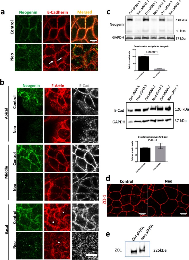
Figure 1.
Neo1 knockdown disrupts adherens junctions and cytoskeletal integrity in Caco-2 cells. (a) Caco-2 cells treated with control or Neo1-siRNA and immunostained for NEO1 (green) and E-Cad (red) showed disruption of the zonula adherens (arrows). The image is a maximum projection image of a z-stack (1 μm). Scale bar-20 μm (b) Caco-2 cells stained for NEO1 (green), F-actin (red) and E-Cad (grey). Images at three apico-basal positions (1 μm, 4 μm and 8 μm) showing disruption of E-Cad localisation and reduction of basal F-Actin rich stress fibres (arrowheads). Scale bar-20 μm (c) Neo1 knockdown in Caco-2 cells was confirmed by Western blot and densitometric analysis. Representative blot with three biological replicates from one experiment and Neogenin blot has been stripped and reprobed for GAPDH. Full length blots for Neogenin and GAPDH are shown in Supplementary Fig. S1. No significant change in E-Cad protein levels after Neo1 knockdown. Each band represents cell lysate proteins from a biological replicate from three independent experiments and E-Cad blot has been stripped and reprobed for GAPDH. Full length blots for E-Cad and GAPDH are shown in Supplementary Fig. S2. (d) Tight junctions were not disrupted after Neo1 knockdown as can be seen with continuous ZO-1 staining (red). Scale bar-20 μm. (e) Western blot for ZO-1 in control and Neo1-siRNA treated cells. Full length blot for ZO-1 is shown in Supplementary Fig. S3.

