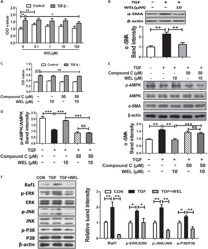
FIGURE 5.

WEL ameliorated TGF-β-induced myofibroblast differentiation partly through Raf1-MAPK signaling pathway and AMPK activation in primary mouse lung fibroblasts (PLFs). The cells were pretreated with compound C (50 μM) or solvent for 1.5 h and subsequently incubated with/without TGF-β1 (10 ng/ml), WEL or solvent for 48 h. (A) The effect of WEL (0.1–100 μM) on PLFs proliferation cells were measured by the MTT assays. (B) The expression of α-SMA in PLFs treated with/without TGF-β1 was detected by Western blotting. (C) The inhibition of WEL or compound C on PLFs proliferation cells were measured by the MTT assays. (D) Expression of p-AMPK/AMPK in PLFs treated with/without TGF-β1 or compound C were determined by Western blotting. (E) The expression of α-SMA in PLFs treated with/without TGF-β1 or compound C were determined by Western blotting. (F) Protein expressions of Raf1, JNK/p-JNK, p38/p-p38, and ERK1/2/p-ERK1/2 in PLFs treated with/without TGF-β1 were detected by Western blotting. Data are presented as mean ± SD (n = 5). ∗p < 0.05, ∗∗p < 0.01, ∗∗∗p < 0.001. NS, non-significant.
