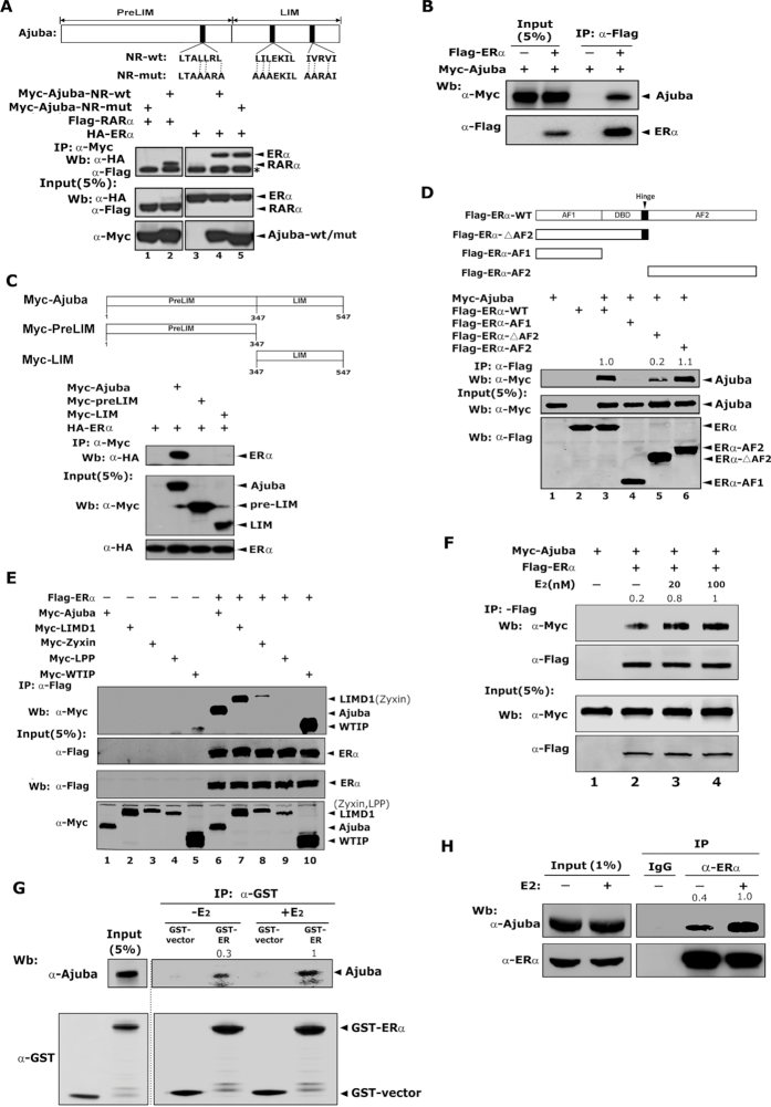
Figure 1.
Ajuba interacts with ERα independent of the conserved NR boxes. (A) The three conserved NR-boxes in Ajuba were shown on the upper panel. Plasmids were transiently transfected into 293T cells and the co-IP assay was carried out with Myc antibody. * IgG-heavy chain. (B) Flag-ERα and Myc-Ajuba plasmids were transfected into 293T cells and co-IP assay was performed by using Flag-M2-beads. (C) The preLIM and LIM regions of Ajuba were illustrated on the upper panel. The full length or truncations of Ajuba were transiently co-transfected into 293T cells together with Flag-ERα plasmid. The co-IP assay was performed by using Flag antibody. (D) The functional domains of ERα were shown on the upper panel. The interaction between full-length or truncations of ERα with Ajuba in 293T cells was detected by co-IP assay and western blotting. The relative amount of co-eluted Myc-Ajuba was semi-quantified by grayscale analysis and the mean values of the three repeats were labeled. (E) The plasmids encoding Ajuba, LIMD1, Zyxin, Wtip and Lpp were respectively co-transfected with Flag-ERα plasmid into 293T cells and co-IP assay was performed. (F) The plasmids encoding Flag-ERα and Myc-Ajuba were transfected into 293T cells, and the resulting cells were cultured in phenol-red free media containing 5% charcoal stripped FBS for 2 days and then treated with E2 (20 or 100 nM) or ethanol for 12 h. The co-IP assay was performed by using Flag-M2 beads. The relative amount of immunoprecipitated Myc-Ajuba was semi-quantified by grayscale analysis and the mean values of the three repeats were labeled. (G) GST-ERα and His-Ajuba proteins were expressed in E. coli BL21, and GST-pulldown assay was performed in the presence of E2 (100 nM) or ethanol. The relative amount of pulled-down His-Ajuba was semi-quantified by grayscale analysis and the mean values of the three repeats were labeled. (H) T47D cells treated with 100 nM E2 or ethanol for 12 h were harvested and co-IP assay was performed by using ERα antibody or IgG control. The relative amount of immunoprecipitated Ajuba was semi-quantified by grayscale analysis and the mean values of the three repeats were labeled.

