Figure 1.
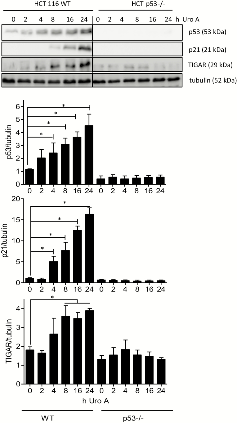
Urolithin A leads to stabilization of p53 and expression of the p53 target genes p21 and TIGAR. WT and p53−/− HCT 116 cells were treated with 30 µM urolithin A for the indicated periods of time. Total cell lysates were then subjected to immunoblot analysis for p53, p21, TIGAR and tubulin as loading control. Representative pictures and compiled densitometric analyses from three independent experiments are depicted (mean ± SD, *P < 0.05, ANOVA, Tukey’s post-test). The bands of WT and p53 knockout lysates originate from one membrane; the line indicates that interjacent bands of no interest (i.e. standard, empty space) were cut out.

