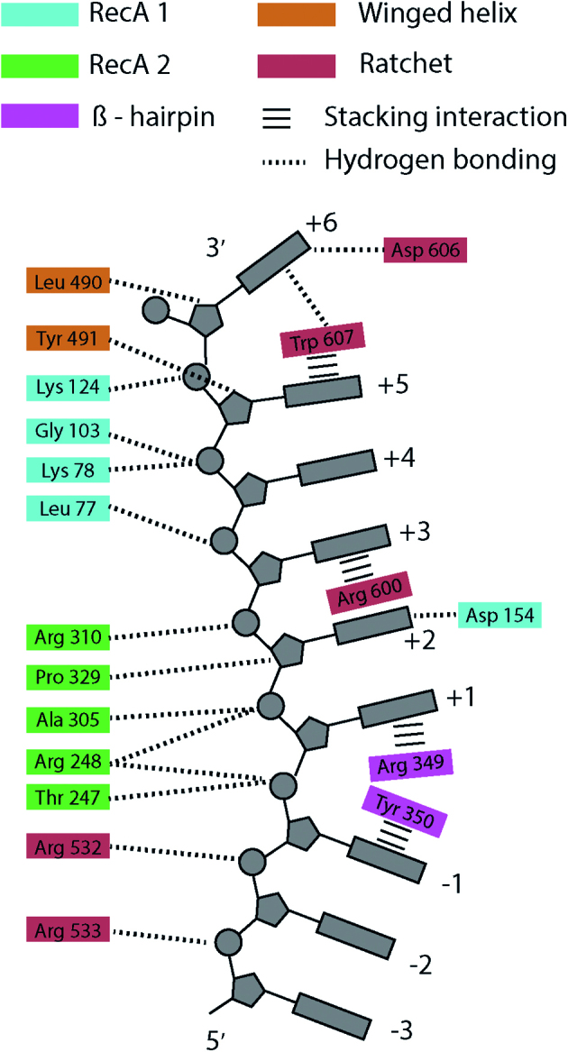
Figure 6.

A cartoon depiction of the suspected contacts between Hel308 amino-acid residues and the template DNA based on comparison of Hel308 from Thermoccocus gammatolerans and the crystal structure of Hel308 from Archaeoglobus fulgidus (23). Hel308 amino acid residues are colored according to the protein domain: RecA domain 1 (blue), RecA domain 2 (green), Beta-hairpin motif (pink), Winged-helix domain 3 (orange), Ratchet domain 4 (red). Dotted lines are hydrogen bonding interactions, and horizontal bars are stacking interactions. The contacts shown on the left interact with the sugar-phosphate backbone, whereas the contacts on the right interact with the bases themselves. The DNA structure is distorted between sites –1 and +1 by the Beta hairpin, and between the +5 and +6 sites by the Ratchet domain. The DNA 3′ end is at the top and the 5′ end at the bottom.
