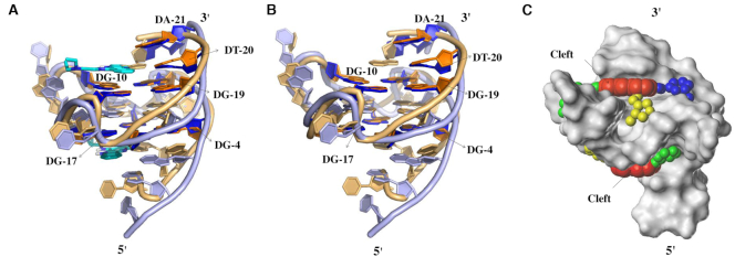
Figure 2.
(A) Cartoon representation of superimposed c-MYC Pu22 G-quadruplex structures in apo state (brown, model 1 in the NMR structure, PDB 1xav) and with quindoline (purple, model 1 in the NMR structure, PDB 2l7v). The two quindoline molecules are shown in cyan. (B) Superimposed structures without ligands. (C) Connolly surface of c-MYC Pu22 G-quadruplex structure from PDB 2l7v. The red spheres highlight the central platform on top of 3′ end G-quartet and at the bottom of 5′ end G-quartet, while green, yellow and blue spheres represent the locations of grooves connecting to the central platform, respectively.

