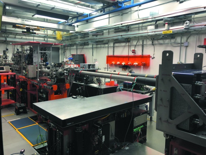
Figure 4.
The MFX hutch as seen from the southeast corner. The unfocused beam enters from the left of the image, and propagates through diagnostics and slits, and continues to the transfocator and further on through more diagnostics and slits. The focused beam exits a diamond window to atmosphere towards the large breadboard of the sample table, seen here empty. To the right is the Rayonix MX340-XFEL detector on the detector mover. Above the table (not seen) is the robot arm, which can also be equipped with another smaller detector. The beam transport pipes for the MEC and CXI instruments can be seen adjacent to the MFX table.

