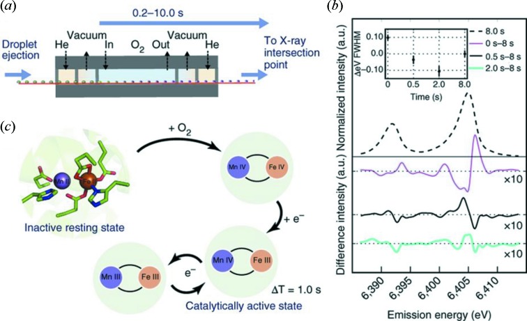
Figure 5.
(a) Schematic of the differentially pumped O2 gas activation setup containing regions of O2 gas and slight negative pressure (vacuum). (b) The known reaction scheme of ribonucleotide reductase R2. (c) Emission spectra of Fe for various O2 exposure times. The inset shows the Kα1 FWHM as a function of exposure time relative to an 8 s exposure. Reproduced with permission from Fuller et al. (2017 ▸).

