Figure 6.
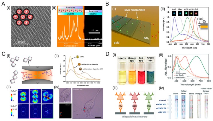
Plasmonic AgNPs for plasmonic nanoantennas and diagnostics. (A) Single-layer AgNP surface-enhanced Raman scattering (SERS) film for a large-scale hot spot. (i) Scanning electron microscopy (SEM) image of a superlattice of 6 nm. AgNPs were used as a homogeneous single-molecule SERS substrate. Illustration shows an interparticle gap for hot spots, which is regulated by the length of a thiolate chain. (ii) Two Raman spectra of single-layered SERS film (left) and quartz surface (right). The enhancement factor was estimated to be larger than 1.2 × 10⁷. Reprinted with permission from [94]. Copyright 2015 American Chemical Society. (B) Metal-film induced plasmon resonance tuning of AgNPs. (i) Schematic illustration of optical scattering spectra of AgNPs on different substrates. (ii) Single AgNP spectra of AgNPs on a silica spacer layer of varying thickness d (nm) on a glass substrate with a 50 nm gold film. The inset is a dark-field image of AgNPs with the corresponding color. The dotted lines represent single particle spectra of AgNPs on a plain glass substrate. Reprinted with permission from [103]. Copyright 2010 American Chemical Society. (C) SERS-based intracellular imaging using alkyne-AgNPs nanoprobes. (i) The structure of colloidal alkyne-AgNP clusters with nano-sized interparticle gaps. (ii) Extinction spectra of the alkyne-AgNPs nanoprobe. The resonance peaks at 400 nm shifted around 520 nm after metal functionalization. (iii) Computational simulation of the far- and near-field optical responses. Intensity distributions of the single particle mode (upper-panels) and the dimer mode (bottom-panels) (iv) Intracellular Raman imaging of a AgNP nanoprobe within the cytoplasmic space of fibroblast. Distinguishable hot spots were highlighted by color-dots related to Raman intensity of the akyne 2045 cm−1 band. Reprinted with permission from [104]. Copyright 2018 Nature Publishing Group. (D) Multiplexed detection with a tunable wavelength of AgNPs. (i) Different colors of AgNPs during a stepwise growth. (ii) Corresponding absorption spectra with varying sizes of AgNPs, such as 30, 41, and 47 nm. (iii) Individual testing of Yellow Fever virus (YFV) NS1 protein, Zaire Ebola virus (ZEBOV) glycoprotein (GP), and Dengue virus (DENV) NS protein using AgNPs. Orange, red, and green AgNPs were conjugated with monoclonal antibodies specific to YFV NS1, ZEBOV GP, and DENV NS, respectively. (iv) Multiplexed detection using different AgNPs-based lateral flow assays. Reprinted with permission from [105]. Copyright 2015 Royal Society of Chemistry.

