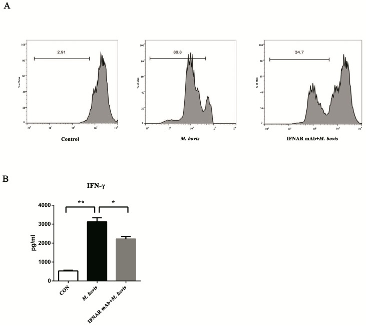
Figure 4.
BMDCs promote T cell activity in the presence of type I interferons. (A) The proliferation of CD4+ T cells was analyzed by flow cytometry after 72 h co-culture. The interferon receptor was treated with neutralizing anti-mouse IFNAR mAb (10 ng/mL) to block and then infected for 24 h with M. bovis (MOI 5). CFSE-stained CD4+ T cells were co-cultured with three different BMDC groups, and the ratios of BMDC:T cells were 1:10 for 72 h. (B) After 72 h co-culture, the culture supernatants were harvested and assessed by ELISA. All data are expressed as mean ± SD. The number above the horizontal bar represents the proliferation rate of CD4+ T cell, (* p < 0.05; ** p < 0.01; n.s.: no statistical significance).

