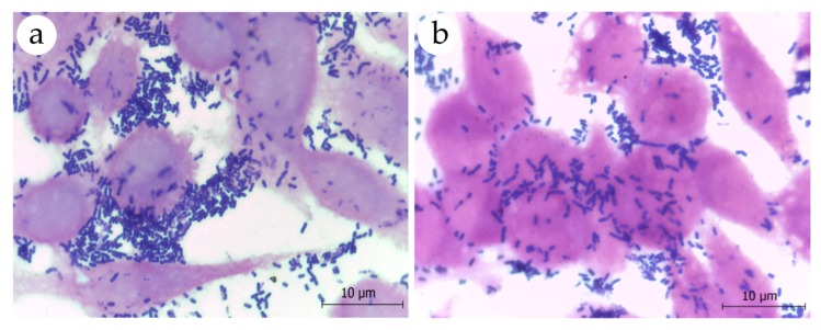
Figure 3.
Microscopic visualization of adhesion assays of (a) L. plantarum ATCC 8014 and (b) L. fermentum ATCC 23271 to HeLa cells after inoculation of approximately 107 CFU of bacterial suspensions. Cell monolayers grown on coverslips were stained by the Gram’s method and examined by light microscopy under a 100× oil immersion objective. Note the presence of numerous gram-positive bacilli adhered to HeLa cells.

