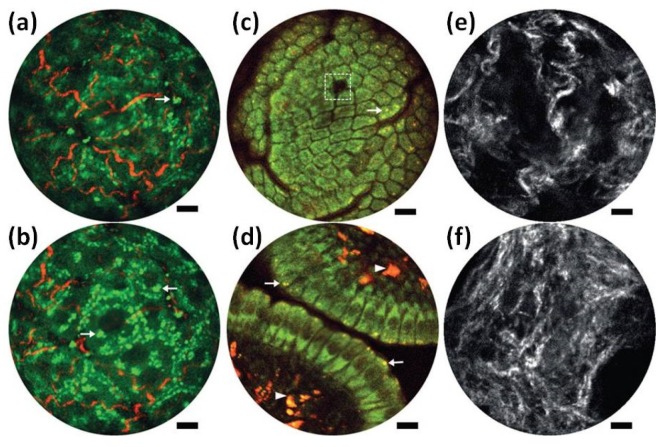
Figure 15.
Endomicroscopy two-photon fluorescence (2PF) and second harmonic generation (SHG) label-free structural imaging. (a,b) Overlay of intrinsic 2PF and SHG ex vivo images of mouse liver. (c,d) Two-photon autofluorescence in vivo images of the mucosa of mouse small intestine. The two detection channels are 417–477 nm for NADH (green) and 496–665 nm for FAD (red). (e,f) SHG images of the cervical collagen fiber network acquired through intact ectocervical epithelium of cervices. Scale bars are 10 μm. Reproduced with permission from [85]; published by Nature, 2017.

