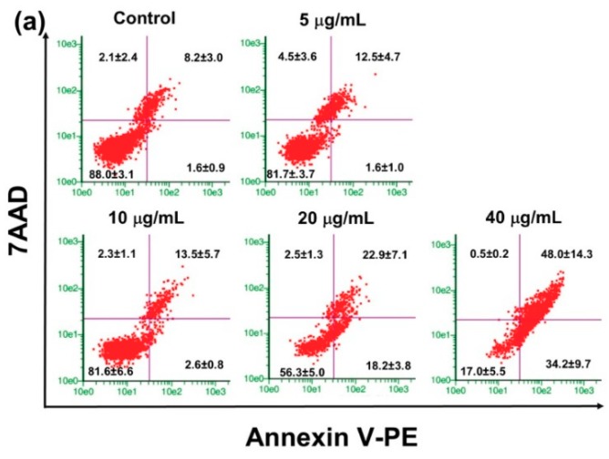
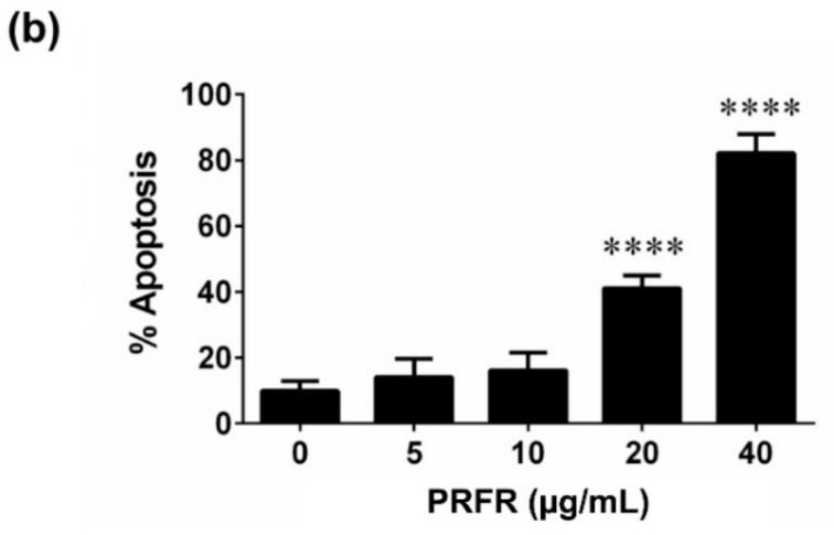
Figure 4.
Effect of PRFR on HepG2 cells apoptosis. The cells were incubated with or without PRFR (0–40 µg/mL) for 48 h. Cell apoptosis was determined using Guava Nexin analysis (a) Annexin V-PE positive cells indicated early apoptosis, while double positive cells indicated late apoptosis. The percentages in early and late apoptosis were summed up; all assays were performed in triplicate in three independent experiments and the mean ± standard deviations are shown as the histogram (b). **** p < 0.0001 versus the control.


