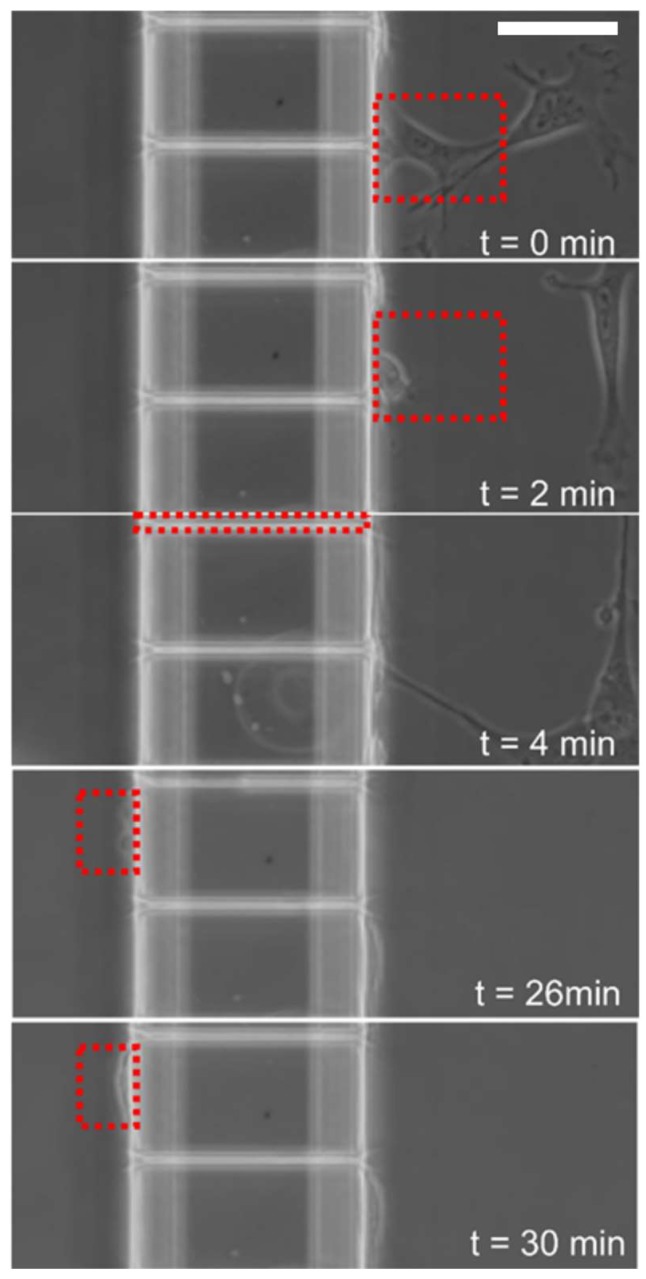
Figure 2.
Time lapse microscopy revealed spontaneous migration of primary myoblast cells across PDMS based microfluidic channels. Phase contrast images of a representative cell at different time intervals traversing a microchannel of 5 μm width and 100 μm length. Red boxes indicate tracking of representative cell from the proximal (right) to distal chamber (left). Horizontal scale bar represents 50 μm.

