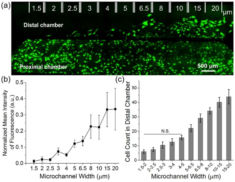
Figure 3.
Migration behavior of primary myoblasts is dependent on the width of microchannels. (a) Representative fluorescent image of cells stained with Calcein AM in the proximal and distal chamber 48 h after seeding. The device consists of microchannel length 100 µm and variable widths (1.5–20 µm) with 6 repeating microchannels per width. (b) Normalized mean intensity of fluorescence in the distal chamber taken for each width from 1.5–20 µm sections. The mean intensity of fluorescence was normalized to the proximal chamber. (c) Quantification of the number of cells in the distal chamber after 48 h from 1.5–20 µm sections. To determine statistically significant differences in the cell count in the distal chamber as a function of microchannel width, a one-way ANOVA with a post hoc Tukey test was performed. Error bars represent standard error of the mean (SEM).

