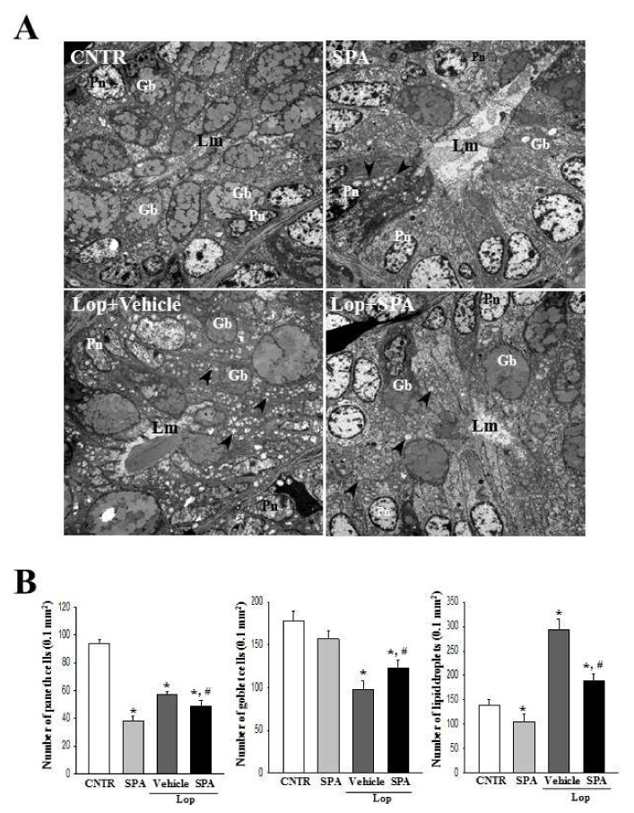
Figure 4.
An ultrastructure image of the colon after SPA administration: (A) The ultrastructure of the crypt in the CNTR, SPA-, Lop + Vehicle- and Lop + SPA-treated groups were viewed by TEM at 4000× magnification. (B) The number of paneth cells, lipid droplets, and goblet cells were measured using Leica Application Suite (Leica Microsystems, Switzerland). The arrow indicates a lipid droplet distributed around the lumen of the crypt. Two to three rats per group were used in the TEM analysis, and each parameter was measured in duplicate in two different slides. The data are reported as the mean ± SD. * indicates p < 0.05 compared to the CNTR group. # indicates p < 0.05 compared to the Lop + Vehicle-treated group. Lm, lumen of crypt; Gb, goblet cells; Pn, paneth cells.

